Positive-Cell Density in Tissue
Counts marker-positive cells in a 2D tissue section and reports positive-cell fraction and density per area.
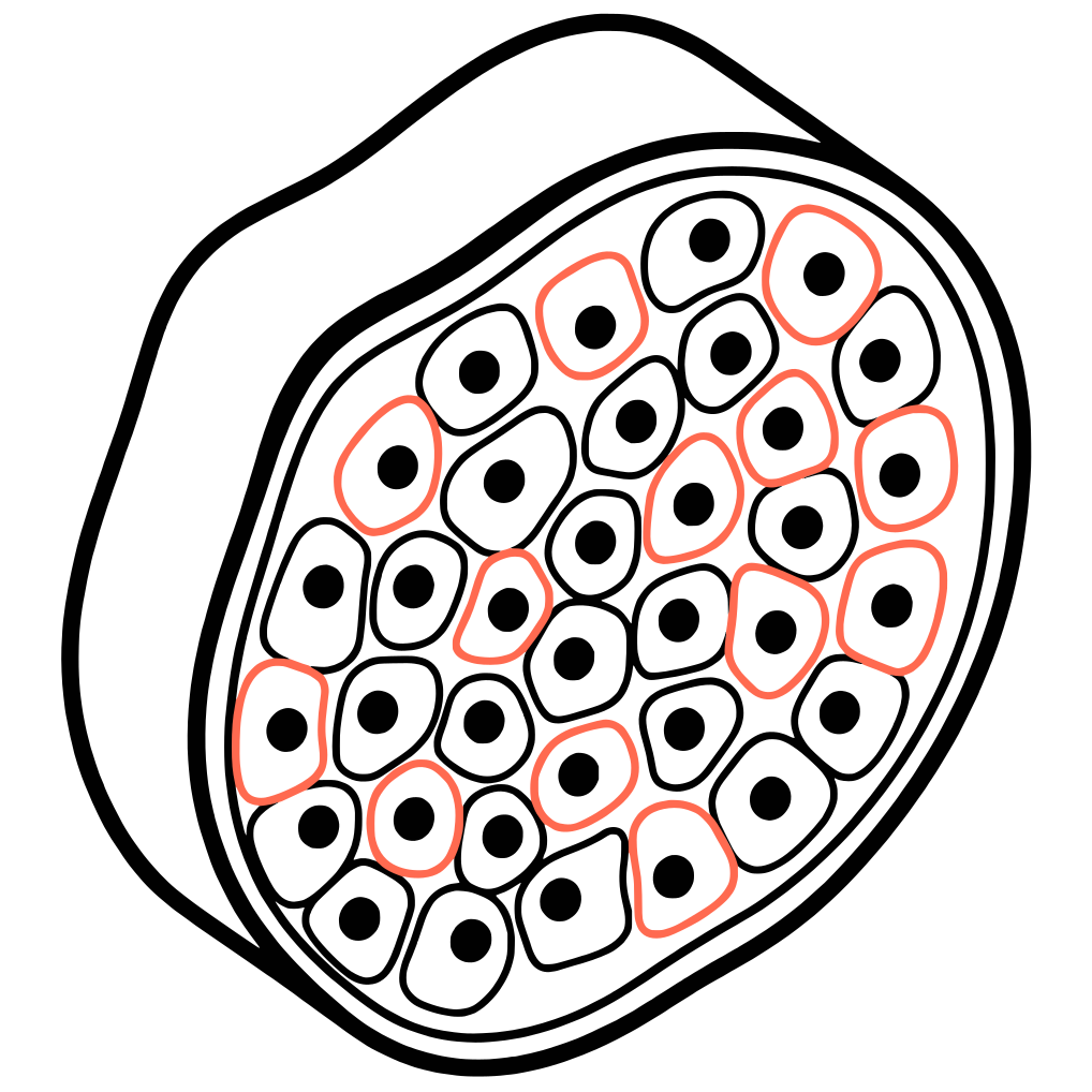
- Ch1: DAPI, Hoechst, or hematoxylin
- Ch2: fluorescent or chromogenic marker
- Optional Ch3-4: cell-type or compartment markers
- Positive-cell count
- % marker-positive cells
- Positive-cell density per unit area
- Marker intensity per cell
Everything you need for Positive-Cell density in tissue
Automated, reproducible, and code-free analysis in the cloud
Cloud-based
No software to install, no GPU required. Run analysis from any browser on any device.
Fully automated
Upload your images and get publication-ready results. No coding or scripting needed.
Reproducible
Every analysis is logged and reproducible. Share protocols with collaborators instantly.
How it runs
Cytely automates the image analysis so you can focus on the science.
Segment nuclei or whole cells
Measure marker intensity per object
Classify positive and negative cells by threshold or gating
Report counts, fractions, and density per area.
Compatibility
Ready to run1
Ki-67 index, cleaved-caspase-3 density, TUNEL-positive density, immune-marker-positive density, lineage-marker positivity in FFPE or frozen sections.Projection-assisted counting may be used for shallow, sparsely labelled stacks where overlap across depth is limited.
Inquire for support
Automated anatomical region segmentation, complex tumour–stroma neighbourhood analysis, 3D tissue density.Projected density remains area-based rather than volumetric and can undercount overlapping cells.
1 Dependent on sample quality
Ready to automate your Positive-Cell density in tissue analysis?
Get started for free or book a demo to see how Cytely works with your data.
Trusted by leading research institutions
Hundreds of researchers use Cytely to accelerate discovery
Used by researchers at












Simple, transparent pricing
Start free. Upgrade when you need more.
Free
For exploration and learning
$0
Free forever
- 1 monthly analysis credit
- Unlimited public datasets
- Community support
Individual
For active researchers
From $299
per month
- 10+ monthly analysis credits
- Image analysis support
- Priority email support
Lab / Team
For collaborating teams
Custom
Contact us
- 5+ seats included
- Shared folders and templates
- Dedicated support
Loved by researchers
Hear from researchers who use Cytely every day.
The data-driven approach that Cytely offers is a dream to an experienced microscopist like me.
This completely removes bias. The price is negligible when you consider the time spent on image analysis.
It's just amazing what can be done today! Cytely does all the work for me!
Cytely makes me 10 times faster. It saves hours on every experiment.

The insight we gained from Cytely was a completely transformative experience. It allowed us to redesign our experiment and finally move the project forward.
Cytely dramatically improved our timelines while enhancing data reliability through automated, unbiased analysis and significantly larger sample sizes.

With Cytely, we instantly analyzed thousands of cells to precisely identify which were transfected, infected, or both. What had been a three-year roadblock was resolved in our first session.
Curious about this assay?
Book a quick call and our team will help you set up your analysis.
Explore more assays


Other assay needs?
Talk to our image analysis specialists. We're adding new assays regularly.