Bacteria Internalization - Salmonella Infected HeLa Cells
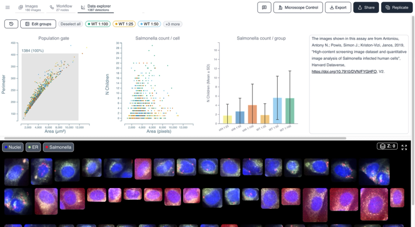
HeLa cells infected with mCherry-labelled Salmonella enterica, imaged by high-content confocal microscopy. This example dataset shows how to detect and quantify intracellular bacteria at the single-cell level, comparing the number of bacteria per host cell across different pathogen concentrations.
See it in action
Watch how this dataset was analyzed end to end in Cytely.
Methodology
Salmonella enterica-infected HeLa cells imaged on a PerkinElmer Opera LX high-content confocal system (40x air objective, NA 0.6). Three fluorescence channels: DAPI (nuclei, 365 nm), ER-tracker (488 nm), and mCherry-Salmonella (561 nm). Cytely segments host cell nuclei and cytoplasm, then detects bacteria as sub-objects within each cell to report the number of bacteria per host cell across pathogen concentrations.
Attribution
Antoniou, A. N., Powis, S. J., & Kriston-Vizi, J. (2018). High-content screening image dataset and quantitative image analysis of Salmonella infected human cells. Harvard Dataverse, V2. https://doi.org/10.7910/DVN/FYGHFO
This dataset is covered by our Data Preservation Policy.

Ready to analyze your own data?
Book a call with our team to see how Cytely turns your microscopy into standardized, reproducible results.