Skin Compartments in a PAS-Stained Section
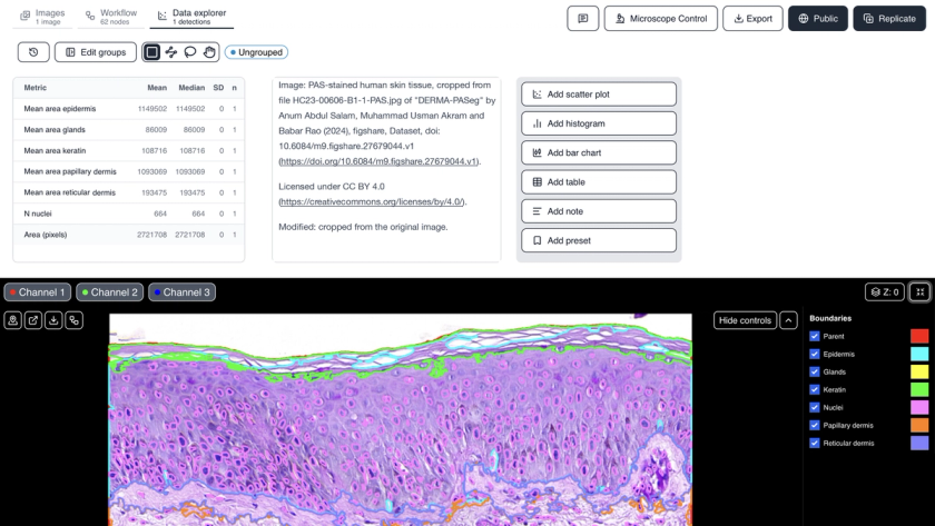
A PAS-stained section of human skin. Cytely outlines keratin, epidermis, papillary dermis, reticular dermis and glands as named regions and detects 664 nuclei across the tissue. Open the shared results to check every outline on the image next to the area of each compartment.
Methodology
One brightfield RGB image, cropped from a whole-slide scan. The tissue is separated from the background, then each compartment is defined by its stain colour: keratin by separating the two stains, the epidermis as the dense nuclei-rich band, and the papillary and reticular dermis and glands by colour range. Size filters, closing and hole filling clean each mask, and the compartments are made mutually exclusive. Nuclei are found on the dark purple signal inside the tissue and split with a watershed; nuclei inside the keratin are removed. Areas are reported in pixels.
Attribution
Image: PAS-stained human skin tissue, cropped from file HC23-00606-B1-1-PAS.jpg of "DERMA-PASeg" by Anum Abdul Salam, Muhammad Usman Akram and Babar Rao (2024), figshare, Dataset, doi: 10.6084/m9.figshare.27679044.v1.

Licensed under CC BY 4.0. Modified: cropped from the original image.
Ready to analyze your own data?
Book a call with our team to see how Cytely turns your microscopy into standardized, reproducible results.