Protein Co-localization - Salmonella and ER Membrane
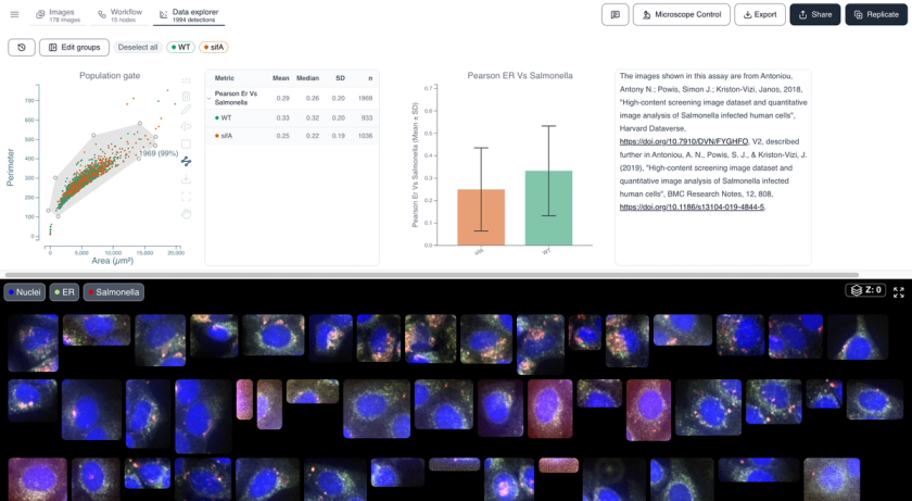
HeLa cells infected with mCherry-labelled Salmonella enterica and stained with an ER-tracker dye, imaged by high-content confocal microscopy. This example dataset shows how to quantify colocalization between intracellular bacteria and the host endoplasmic reticulum, comparing wild-type and isogenic ΔsifA mutant strains.
See it in action
Watch how this dataset was analyzed end to end in Cytely.
Methodology
Salmonella enterica-infected HeLa cells imaged on a PerkinElmer Opera LX high-content confocal system (40x air objective, NA 0.6). Three fluorescence channels: DAPI (nuclei, 365 nm), ER-tracker (488 nm), and mCherry-Salmonella (561 nm). Cytely segments host cell nuclei and cytoplasm, then computes a per-cell Pearson correlation between the Salmonella (mCherry) and ER-tracker channels, comparing wild-type versus isogenic ΔsifA mutant infection.
Attribution
Antoniou, A. N., Powis, S. J., & Kriston-Vizi, J. (2018). High-content screening image dataset and quantitative image analysis of Salmonella infected human cells. Harvard Dataverse, V2. https://doi.org/10.7910/DVN/FYGHFO
This dataset is covered by our Data Preservation Policy.

Ready to analyze your own data?
Book a call with our team to see how Cytely turns your microscopy into standardized, reproducible results.