Blood Cell Analysis - Neutrophils
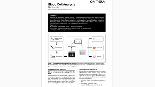
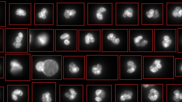
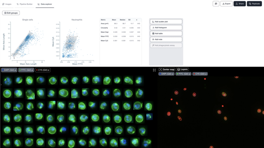
백혈구, 혈소판, 적혈구(RBC)가 혼합된 전혈 시료 내 세포 집단을 분석합니다. 이 데이터셋은 산점도(scatter plot)와 직관적인 게이팅을 통해 다양한 혈액 세포 유형을 단일 세포 수준에서 신속하게 식별하고 정량화하는 과정을 보여줍니다. 이질적인 시료로부터 호중구(57%), 혈소판(11%), 적혈구(12%)를 성공적으로 식별합니다.
방법론
건강한 지원자에게서 정맥천자로 말초혈액을 채취했습니다. 호중구는 Ficoll-Paque를 사용한 밀도 구배 원심분리로 분리했습니다. 세포는 CellMask™ Green Plasma Membrane Stain(2 µM), CellTracker™ Deep Red(1 µM), Hoechst 33342 핵 염색(5 µg/mL)으로 염색했습니다.
사용 예시
Visualizing heterogeneity across mixed cell populations. Identifying populations (neutrophils, platelets, RBCs) using scatter plots. Gating to isolate neutrophil subsets. Quality control of blood cell preparations.
기술적 참고 사항
Three fluorescent channels used: Hoechst 33342 (350/461 nm) for nuclei, CellMask Green (522/535 nm) for membrane, and CellTracker Deep Red (630/650 nm) for cytoplasm. Images acquired using Nikon TI2 widefield microscope.