手法
HeLa細胞を8ウェルibidiスライドで培養し、4% PFAで15分間固定しました。細胞はHoechst 33342(核染色)およびPhalloidin-Alexa Fluor 488(アクチン細胞骨格)で染色しました。画像はNikon TI2広視野顕微鏡でDICを用いて取得しました。
使用例
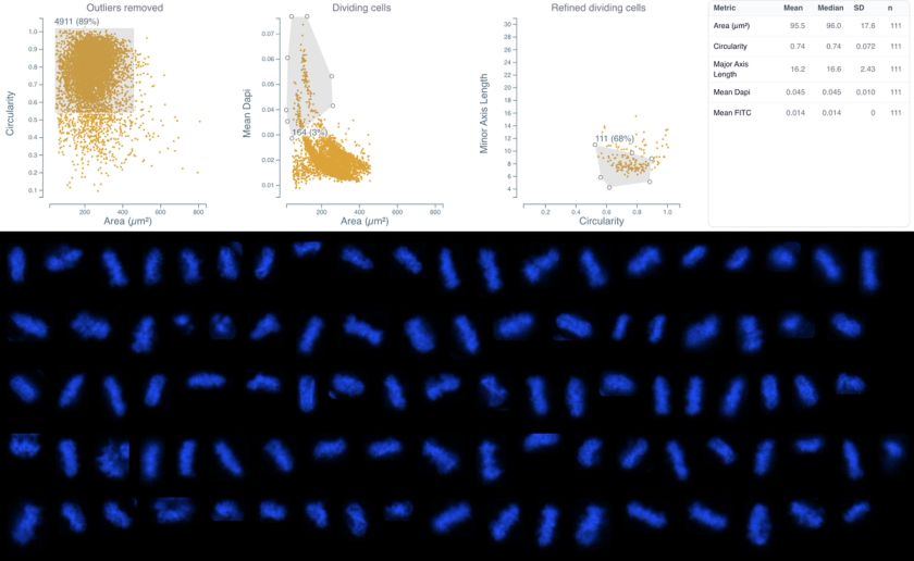
Complex cell cycle analysis without specialized dyes (e.g., EdU, BrdU). Retrospective analysis of existing datasets. Sequential gating to isolate mitotic cells with high purity. Morphological profiling of cell cycle stages.
技術的な注記
Key morphological features used: Area, Circularity, Mean DAPI Intensity, Major Axis Length, Minor Axis Length. Final gating based on nuclear elongation (Major vs. Minor Axis Length) was critical for removing contaminating interphase cells. Distribution: G0/G1 (65%), S/G2 (23%), Mitosis (3%), Other/Debris (9%).
このデータセットで使用されているアッセイ

Cell Morphology & Phenotypic Profiling
細胞形態・表現型プロファイリング
複数のマーカーにわたって個々の細胞のサイズ、形状、蛍光強度を測定します。
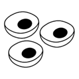
Cell Growth, Survival & Fitness
細胞数計測・生存率
総細胞数を計測し、マーカー強度に基づいて生細胞か死細胞かを分類します。毒性試験や用量反応実験において最も基本的な評価指標を提供します。
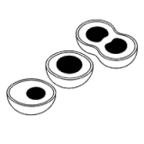
Cell Growth, Survival & Fitness
細胞周期解析
細胞の形態とDNA染色強度を用いて細胞をG0/G1、S/G2、M期に分類し、フローサイトメトリーとは異なり空間的コンテキストを保持します。