Cell Cycle Analysis - Mitotic Cells in HeLa Cultures
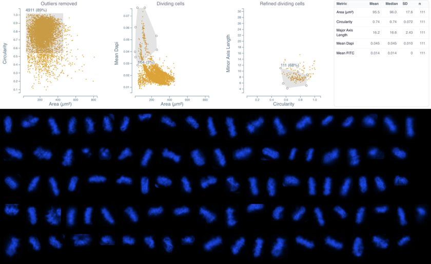
표준 핵 염색만을 사용하여 형태학 기반 분석으로 HeLa 세포의 세포주기 단계를 분류합니다. 이 데이터셋은 특수 염료 없이 핵 형태만으로 세포주기 시기(G0/G1, S/G2, M)를 식별하는 과정을 보여줍니다. 핵 특성에 기반한 순차적 게이팅을 통해 약 5,500개의 전체 세포 중 164개의 유사분열(mitotic) 세포(3%)를 성공적으로 분리했습니다.
방법론
HeLa 세포를 8웰 ibidi 슬라이드에서 배양하고 4% PFA로 15분간 고정했습니다. 세포는 Hoechst 33342(핵 염색)와 Phalloidin-Alexa Fluor 488(액틴 세포골격)로 염색했습니다. 영상은 DIC를 갖춘 Nikon TI2 광시야(widefield) 현미경으로 획득했습니다.
사용 예시
Complex cell cycle analysis without specialized dyes (e.g., EdU, BrdU). Retrospective analysis of existing datasets. Sequential gating to isolate mitotic cells with high purity. Morphological profiling of cell cycle stages.
기술적 참고 사항
Key morphological features used: Area, Circularity, Mean DAPI Intensity, Major Axis Length, Minor Axis Length. Final gating based on nuclear elongation (Major vs. Minor Axis Length) was critical for removing contaminating interphase cells. Distribution: G0/G1 (65%), S/G2 (23%), Mitosis (3%), Other/Debris (9%).
이 데이터셋에 사용된 분석

Cell Morphology & Phenotypic Profiling
세포 형태 및 표현형 프로파일링
여러 마커에 걸쳐 개별 세포의 크기, 형태, 형광 강도를 측정합니다.

Cell Growth, Survival & Fitness
세포 계수 및 생존율
전체 세포 수를 세고 마커 강도를 기반으로 세포를 생존 또는 사멸로 분류하여, 독성 및 용량-반응 실험을 위한 가장 기본적인 판독값을 제공합니다.

Cell Growth, Survival & Fitness
세포 주기 분석
세포 형태와 DNA 염색 강도를 사용하여 세포를 G0/G1, S/G2, M기로 분류하며, 유세포 분석과 달리 공간적 맥락을 보존합니다.